A pipeline to study hominoid brain evolution

Friederici et al 2024

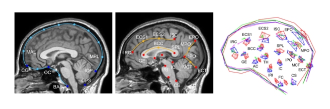
A large team led by Angela Friederici has now published a methodological article to describe a pipeline to investigate brain evolution, integrating anatomical and behavioral information from living and extinct species. To analyze correlations between brain structure and function, post-mortem brain imaging and histology in non-human primates are matched with (pre-mortem) behavioral data. Such correlations can be extrapolated to fossil endocasts. In living primates, shape analysis is employed to localize endocranial surface features, then matched with the sulcal pattern of the corresponding brain. Adult skulls are micro-CT scanned with a resolution of 90 μm. Results are, indeed, promising and exciting, revealing a very good correspondence between endocranial imprints and cortical elements. According to the authors, “almost all brain sulci leave unambiguous impressions in the endocranial cavity”. Major differences between endocranial and brain form are due to brain shrinking (mostly in the vertical dimension) after extraction, and removal of the connective tissues and cerebrospinal fluid. I guess the absence of blood pressure is another crucial factor. In the paper, language is finally used as an example to discuss how this pipeline can be employed to investigate cognitive functions that are key features in human evolution.

The large number of people, institutions, and methods necessary to handle this pipeline reminds us that approaches like this one are, indeed, very difficult to organize. Technical outputs, methodological issues, and their final interpretations represent an intricate network of variables and parameters, which require much caution when coming to the final inferences and conclusions. Nonetheless, the publication of this article also suggests that such comprehensive and efficient approaches are, at present, already available. And this is definitely the very good news.

This article represents a very useful and efficient tutorial on current comparative neuroanatomy, in particular when dealing with traditional aspects associated with evolutionary anthropology. We must anyway remark (once more) two relevant points. First, in this field, comparative approaches with humans and apes are essential. However, we should not forget that living apes are derived taxa, not ancestral ones. Furthermore, they are specialized for a kind of environment profoundly different from the one in which humans evolved. Their striking differences in cranial architecture add one more crucial problem, when they are used as models in human paleoneurology and, in general, for human brain or cognitive evolution. The second point deals with the fact that we keep focusing on human evolution as the main target of research. This is undeniably a priority, no doubt. But it should not be the only target. Apes biology and evolution are also amazing topics, to which we should probably pay more attention. And, in this case, we should probably avoid taking humans as models when investigating their peculiar derived traits.

Subcortical invariants

Amano et al 2023 - Invariant brain shape

I have recently read this article by Amano and colleagues on subcortical shape variations in humans and great apes. When compared with cortical morphology, these deep regions display a minor degree of variability. The study is aimed at considering whether these regions can be estimated in fossil species, which seems likely, taking into account their apparently conserved morphology. Nonetheless, we should also remark that, beyond macroscopic shape differences, many of these areas show specializations (in functions, connectivity or cytoarchitecture) that cannot be detected on the basis of gross spatial organization. Human specializations in deep brain regions include, for example, functions associated with language and emotions. In the former case, it has been even proposed that such deep changes may influence the general brain morphology. Actually, according to the results of this new paper, even among living apes there are anyway shape differences that, albeit not outstanding, are significant. Furthermore, beyond evolutionary or functional issues, we must consider that these deep regions suffer important topological constraints, because of their physical position, embedded into the core of the brain mass and on the top of the cranial base. In 2007, Nathan Jeffery and I published a very early analysis on this same topic, remarking the influence of the cranial base, a general allometric trend, and a curious departure of gibbons. This new article by Amano and colleagues is definitely welcome, calling the attention on a stimulating challenge in paleoneurology: the analysis of cerebral elements hidden in the deep black box of an endocast. Although largely based on inferences and indirect evidence, this is an issue that should not be neglected at all.

Asymmetries, encore …

I had missed this further study on endocranial asymmetries. The issue is central in brain evolution because hemispheric asymmetries has been since ever interpreted as a specialized human feature. Endocranial asymmetries, used as proxy for brain asymmetries, are then generally used to investigate this character in fossils. Different scholars have supported different conclusions on this matter. It has been suggested that hemispheric asymmetries are specific of humans (with relevant evolutionary implications), that the difference is only a matter of degree (humans would hence have just more asymmetries than other primates, but of the same nature), or that humans are more asymmetric only because of a larger brain size (allometry). These alternatives are then mixed with causal hypotheses involving genetic aspects, structural adjustments, or neural plasticity. As for many other paleo topics, anyone has a personal recipe on how we can interpret these phylogenetic similarities and differences. And, importantly, on how to measure them. The issue of measurement is not secondary, because we are dealing with smooth, blurred, and tiny morphometric features, not easy to detect (and to quantify) at all. In this new study, Simon Neubauer and colleagues employed an elegant and complex set of mathematical adjustments to compare both the kind and degree of endocranial asymmetries in humans and apes. According to their results, chimps are less asymmetric than humans, but orangs and gorillas display a human-like pattern of asymmetries. Or, we should say, an ape-like pattern of petalias (the gross right-left endocranial asymmetries). These differences are due to size variation only to a limited extent, suggesting that asymmetries could be a functional thing, and not an allometric consequence of large brain volumes. The occipital asymmetry stands as the most noticeable one, in both humans and apes. With all this in mind, it is anyway useful to recall two main points. First, when dealing with the human genus (Homo), all human species are known to display the modern (Homo sapiens) pattern. So, whatever the conclusions on this topic, it does not add to major questions internal to the evolution of our own species. The fact that the modern pattern of endocranial asymmetries is shared within all the human genus is something known since long ago, although many scholars keep on discussing this aspect in any new fossil specimens, as it would be of any interest (I guess that they found the topic academically “sexy” for media and colleagues, but it is actually like one would stress, at any new fossil hominid discovery, that the individual had two legs). Second, it is necessary to remember that we still don’t know what is the biological relationship between those macroscopic skull asymmetries and the corresponding histological brain asymmetries. Those endocranial asymmetries are due to differences in brain mass, neurons, neural connections, blood, cerebrospinal fluid, cranial constraints, genetic programs, biomechanical tensions, and a large series of morphogenetic factors. To date, the underlying mechanisms generating those subtle differences are not known, and a direct association between endocast asymmetries and brain asymmetries may be a risky (and hasty) speculation.

Humans, chimps, and brain size

Amélie Beaudet and colleagues have now published a new review on the evolution of the modern human brain. They introduce many traditional issues in paleoneurology, including frontal lobe evolution, asymmetries, lunate sulcus, brain growth, and brain shape. They also provide a detailed discussion of the information we have on the evolution of brain size. Studies on this topic are frequently biased by statistical or taxonomic problems, because of the intrinsic limitations of the fossil record. Actually, any model (gradual, random, punctuated, etc.) can be supported by the few and scattered data, generating disagreements and debates. Isaac Asimov said “Where any answer is possible, all answers are meaningless”. In this paper, they describe their own approach, trying to deal with such limitations. They support a gradualist perspective, although with some discontinuities within some clades. The review strictly deals with brain evolution, but I really appreciate the taxonomic considerations at the beginning of the article, defending their reasons to include humans and apes in one single family. I belong to the opposite faction, namely to the resisting supporters of two distinct families for this group, with the term hominids restricted to humans and (probably) australopiths. Firstly, because I think that taxonomy should not try to trace phylogeny too strictly, constrained and forced by cladistic schemes. The real phylogeny is unknown (we use genes as a proxy, which is but an estimation, with pros and cons), and the phylogenetic hypotheses are frequently changing. Instead, a taxonomy based on the whole biological model (that includes anatomy, physiology and so on) is more stable and, importantly, can add more information on the actual evolutionary, zoological and ecological organization and role of a group of species. Secondly, because I think that differences are the great value of evolution, and taxonomy should acknowledge such differences. In this case, we must admit that our lineage is particularly dissimilar from all the other apes. This does not mean that we are better, but surely much different, and taxonomy should take into account the importance of such outstanding changes. Many anthropologists give all these taxonomical issues for granted, using one label or another just by repeating or copy-pasting what they hear around, generally following a mainstream without a personal or competent opinion. But passively repeating statements is proper of dogmas and mantras, something that should be left out of science.  That’s why I really appreciate that, in this article, Amélie and her coauthors take a clear position, explaining their reasons.

***

More on humans and chimps can be found in this other recent review on human paleoneurology. For Spanish readers, here a provocative dissemination article on humans and apes, and another one on the immense value of diversity.

Shaping cortical evolution

Happy 2019 to everybody! To begin with this new year, here a new review on human paleoneurology, published in Journal of Comparative Neurology. Some conceptual and methodological issues in functional craniology, digital anatomy and computed morphometrics are introduced and discussed. The case-study on parietal evolution is also briefly summarized, with special attention to connectivity. Nonetheless, more specifically, the review points to theoretical and practical limitations of the field. Living species can provide information on the product of evolution, while fossils are necessary to provide information on the process. In the former case (extant species) we can rely on more comprehensive biological analyses, but results concern the final result of the process, not the process itself. In the latter case (extinct species) we can investigate directly the process, but samples are generally not representative neither at biological nor at statistical level. This dual framework is often not properly acknowledged, confounding taxonomy (the product) with phylogeny (the process). When samples and information are analyzed without these cautions in mind, conclusions can generate misleading hybrid perspectives. From the one hand, living species (monkeys and apes in anthropology and evolutionary neuroscience) are still frequenlty misinterpreted as primitive human ancestors. At the same time, scattered and descriptive information on individual and fragmented fossils are generalized to propose broad and inclusive theories. Both aspects are, scientifically speaking, crucial weaknesses, generating instability and unreliability within the field.

Another issue concerns the Homo-centric perspective that still contaminates evolutionary neuroanatomy and evolutionary anthropology. Apart from generating a deformed evolutionary scenario, anthropocentric views demote attention towards the other primates. Apes are generally used to “shed light on human evolution”. But living apes are not ancestral to humans. They could be bad models to understand our evolution, as we humans are probably bad models to understand their own one. They have their own specialized traits, which merit attention. In fact, apes are themselves an exceptional zoological case study. Anthropology is interesting, but apeology is interesting too. In cognitive terms, for example, apes could have capacities that we have never evolved. Finally, it can be also worth nothing that, charmed in searching for “what makes us humans”, we are neglecting “what makes us primates”. Because these latter features are associated with instincts, emotions, and cognitive constraints, they seriously deserve attention. Mostly when recognizing that they often deal with our social aspects, and with their consequences.

Integrated paleoneurology

Zollikofer et al 2016Together with the recent article on modern vs Neandertal endocranial ontogeny, the team coordinated by Christoph Zollikofer has now published also a large and comprehensive study on endocranial ontogeny in humans and apes. The paper focuses on a specific question: to what extent endocranial differences are due to brain differences, and to what extent they are due to cranial constraints? Definitely, this is a key-paper in paleoneurology. They considered the integration between and within the main cranial districts to evaluate the influence on brain shape of two major cranial effects: spatial packing and facial orientation. Their analyses suggest that endocranial differences between humans and apes, as well as differences among apes, are the result of all those factors, the cerebral and the cranial ones. Therefore, the endocranial form is due to a complex admixture of specific brain differences (already present at birth) and cranial constraints. Comparisons among endocranial ontogenetic patterns of living hominoids, among adult fossil specimens, and among different neuroanatomical aspects of living species, can give different results, suggesting that the relationships between anatomical, morphological, and cytological elements is far from being understood. In my opinion, a limit of many shape analyses in general concerns the use of surface semi-landmarks to analyze brain geometry. Surface landmarks are necessary because of the lack of good anatomical references on the endocasts. Unfortunately, they can’t take into account the contribution of distinct cerebral areas, and as a consequence they consider brain morphology as a single homogeneous surface. The identification of boundaries or distinct and independent elements within this surface might seriously influence the multivariate output. I am particularly interested in the analysis of the parietal districts. When using surface landmarks the analysis of the parietal surface may give different (and sometimes contrasting) results. Hence, we may wonder whether the observed parietal variations are the result of brain differences (cortical expansion/reduction) or of geometry (bulging and flexion). Nonetheless, previous morphological studies based on cortical landmarks suggest that modern humans show an actual (absolute and relative) increase not only of the parietal “surface”, but also and specifically of the parietal “lobe”, when compared with extinct hominids or with living chimps. The localization of anatomical boundaries on endocasts may be difficult, although those results have been replicated on different samples. The identification of anatomical landmarks in living species is, in contrast, definitely more reliable. Therefore, whatever the result of a global surface analysis of the whole endocranium, we should not forget that comparisons of specific areas are suggesting a differential contribution of distinct brain components.

Hominoids

Scott et al 2014After shape analysis of the endocranial growth and development in modern humans, chimps, and Neandertals, the team from the Max Planck Institute has published a study on apes endocranial ontogeny. In their former articles they evidenced a shared trajectory of form change in humans and chimps. The only exception is the “globularization stage” in modern humans, an early postnatal stage associated with parietal and cerebellar enlargement. This study now includes also gorillas, orangs, and gibbons, confirming that after eruption of the deciduous dentition all hominoids share a similar pattern of form variation. Differences among species are largely a matter of degree of change, but within a shared set of rules. This implies that most of the observed differences among their endocranial forms take place before, in prenatal stages.