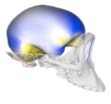
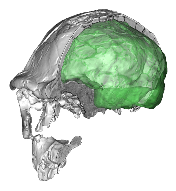
 Together with the recent article on modern vs Neandertal endocranial ontogeny, the team coordinated by Christoph Zollikofer has now published also a large and comprehensive study on endocranial ontogeny in humans and apes. The paper focuses on a specific question: to what extent endocranial differences are due to brain differences, and to what extent they are due to cranial constraints? Definitely, this is a key-paper in paleoneurology. They considered the integration between and within the main cranial districts to evaluate the influence on brain shape of two major cranial effects: spatial packing and facial orientation. Their analyses suggest that endocranial differences between humans and apes, as well as differences among apes, are the result of all those factors, the cerebral and the cranial ones. Therefore, the endocranial form is due to a complex admixture of specific brain differences (already present at birth) and cranial constraints. Comparisons among endocranial ontogenetic patterns of living hominoids, among adult fossil specimens, and among different neuroanatomical aspects of living species, can give different results, suggesting that the relationships between anatomical, morphological, and cytological elements is far from being understood. In my opinion, a limit of many shape analyses in general concerns the use of surface semi-landmarks to analyze brain geometry. Surface landmarks are necessary because of the lack of good anatomical references on the endocasts. Unfortunately, they can’t take into account the contribution of distinct cerebral areas, and as a consequence they consider brain morphology as a single homogeneous surface. The identification of boundaries or distinct and independent elements within this surface might seriously influence the multivariate output. I am particularly interested in the analysis of the parietal districts. When using surface landmarks the analysis of the parietal surface may give different (and sometimes contrasting) results. Hence, we may wonder whether the observed parietal variations are the result of brain differences (cortical expansion/reduction) or of geometry (bulging and flexion). Nonetheless, previous morphological studies based on cortical landmarks suggest that modern humans show an actual (absolute and relative) increase not only of the parietal “surface”, but also and specifically of the parietal “lobe”, when compared with extinct hominids or with living chimps. The localization of anatomical boundaries on endocasts may be difficult, although those results have been replicated on different samples. The identification of anatomical landmarks in living species is, in contrast, definitely more reliable. Therefore, whatever the result of a global surface analysis of the whole endocranium, we should not forget that comparisons of specific areas are suggesting a differential contribution of distinct brain components.
